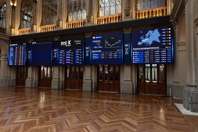
Exploring Cardano: Inner Workings and Advantages of this Cryptocurrency
 Seville.- Economy.- Innova.- STSA inaugurates its new painting and sealing hangar in San Pablo, for 18 million
 Innova.- More than 300 volunteers join the Andalucía Compromiso Digital network in one month to facilitate access to ICT
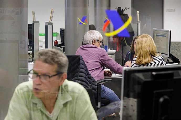
 Innova.-AMP.- Ayesa acquires 51% of Sadiel, which will create new technological engineering products and expand markets
 Abascal (Vox) criticizes that Sánchez is "victimizing" himself and calls for elections after his possible resignation
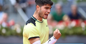 Carlos Alcaraz reaches the round of 16 in Madrid without breaking a sweat
 Some 5,000 people demonstrate in front of Congress for democracy, hours before Sánchez's decision
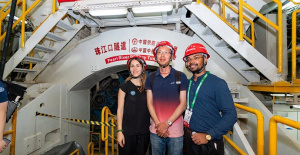 STATEMENT: Intelligent systems used in the construction of the deepest underwater tunnel in China
 How Blockchain in being used to shape the future
 Not just BTC and ETH: Here Are Some More Interesting Coins Worth Focusing on
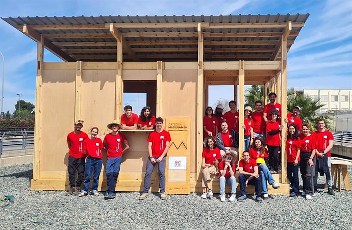
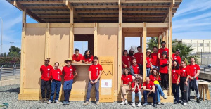 UPV students build a prototype of a wooden house to move to Equatorial Guinea
 The UA opens the call for the Impulso 2024 Awards for the best innovative business initiatives
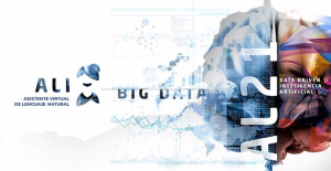 ALI, virtual assistant from Alicante, internationally recognized by the OECD
 Retrópolis brings the golden age of video games and computing to the UPV
 A million people demonstrate in France against Macron's pension reform
 Russia launches several missiles against "critical infrastructure" in the city of Zaporizhia
 A "procession" remembers the dead of the Calabria shipwreck as bodies continue to wash up on the shore
 Prison sentences handed down for three prominent Hong Kong pro-democracy activists
 ETH continues to leave trading platforms, Ethereum balance on exchanges lowest in 3 years
 Investors invest $450 million in Consensys, Ethereum incubator now valued at $7 billion
 Alchemy Integrates Ethereum L2 Product Starknet to Enhance Web3 Scalability at a Price 100x Lower Than L1 Fees
 Mining Report: Bitcoin's Electricity Consumption Declines by 25% in Q1 2022
 Oil-to-Bitcoin Mining Firm Crusoe Energy Systems Raised $505 Million
 Microbt reveals the latest Bitcoin mining rigs -- Machines produce up to 126 TH/s with custom 5nm chip design
 Bitcoin's Mining Difficulty Hits a Lifetime High, With More Than 90% of BTC Supply Issued
 The Biggest Movers are Near, EOS, and RUNE during Friday's Selloff
 Global Markets Spooked by a Hawkish Fed and Covid, Stocks and Crypto Gain After Musk Buys Twitter
 Bitso to offset carbon emissions from the Trading Platform's ERC20, ETH, and BTC Transactions
 Draftkings Announces 2022 College Hoops NFT Selection for March Madness
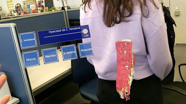
VALENCIA, Apr. 27 (EUROPA PRESS) -
Research staff from the Mixed Biomedical Imaging Unit of the Fisabio Foundation and the Príncipe Felipe Research Center (CIPF), led by the researcher María de la Iglesia, have developed a prototype that uses magnetic resonance imaging (MRI) of the lumbar spine and virtual reality glasses to assist in lumbar spine surgeries.
The prototype is being developed with the Hololens device (holographic device) that simulates a 3D model of the patient's spine in the same position as the real spine. With this, surgeons will be able to see the entire anatomy of the lumbar spine prior to surgical intervention.
The model, which is being developed by the researcher at the Fisabio-CIPF Mixed Biomedical Imaging Unit Kate Kardash, simulates the spine in a virtual way, with realistic movements and in different positions, where the different points that will be intervened during the procedure can be marked. surgery.
The spinal models generated with the program have been created through magnetic resonance imaging, where the spine has been previously segmented --using Artificial Intelligence-- with more than eleven identified structures of the lumbar spine. All these images have been extracted from the MIDAS (Massive Image Data Anatomy Spine) project.
The researcher from the Fisabio-CIPF Mixed Biomedical Imaging Unit and the University of Navarra Clinic, Julio Domenech, has stated that "unlike reconstructions based on computed tomography (CT), which only allow bone reconstruction, lumbar MRI images give very good resolution to identify other structures such as nerves, spinal cord, discs, muscle and ligaments in the reconstructed 3D image."
On the other hand, the research staff will continue to improve the tool to create a connection of multiple glasses that can be used by different specialists of the medical team.
For her part, María de la Iglesia, principal investigator of the Fisabio-CIPF Mixed Biomedical Imaging Unit, explained that "the program is still in the prototype phase, since all its features have been tested with virtual models, but not in real samples.
In addition, its future development could be applied not only in spinal surgeries, but in any part of the body or tissues that could help healthcare personnel in the diagnosis and treatment of certain injuries in clinical practice."
"The model has two applications that can be transferred to clinical use in patients. On the one hand, it makes it easier for the surgeon to plan the surgery in advance, and on the other, it can be used intraoperatively, which will make it possible to more accurately identify anatomical structures, so that increases the precision of surgical gestures and access to structures not visible with the ordinary technique", added Julio Domenech.