Exploring Cardano: Inner Workings and Advantages of this Cryptocurrency
 Seville.- Economy.- Innova.- STSA inaugurates its new painting and sealing hangar in San Pablo, for 18 million
 Innova.- More than 300 volunteers join the Andalucía Compromiso Digital network in one month to facilitate access to ICT
 Innova.-AMP.- Ayesa acquires 51% of Sadiel, which will create new technological engineering products and expand markets
 STATEMENT: ELFBAR and LOST MARY reveal progress in the fight against illicit vapers (1)
 STATEMENT: ELFBAR and LOST MARY reveal progress in the fight against illicit vapers (2)
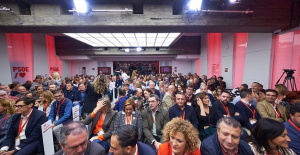 The PSOE is holding a Federal Committee this Saturday that will serve to close ranks with Sánchez so that he does not resign
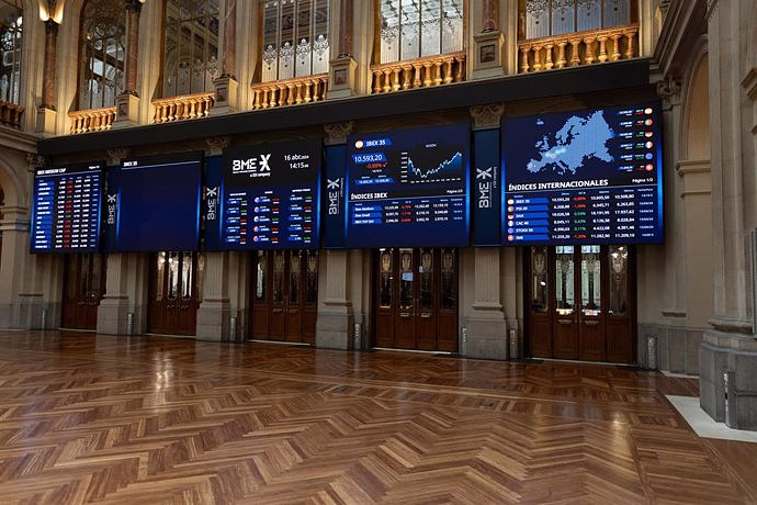
 The Ibex 35 closes the week at its highest since 2015 and is already looking at 11,200
 How Blockchain in being used to shape the future
 Not just BTC and ETH: Here Are Some More Interesting Coins Worth Focusing on
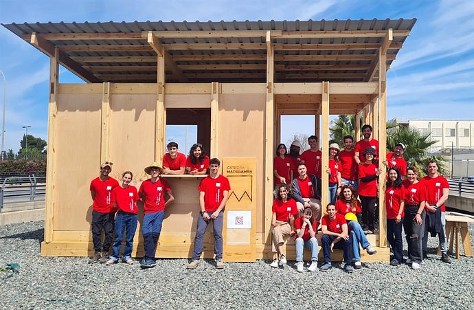
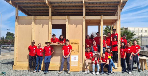 UPV students build a prototype of a wooden house to move to Equatorial Guinea
 The UA opens the call for the Impulso 2024 Awards for the best innovative business initiatives
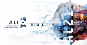 ALI, virtual assistant from Alicante, internationally recognized by the OECD
 Retrópolis brings the golden age of video games and computing to the UPV
 A million people demonstrate in France against Macron's pension reform
 Russia launches several missiles against "critical infrastructure" in the city of Zaporizhia
 A "procession" remembers the dead of the Calabria shipwreck as bodies continue to wash up on the shore
 Prison sentences handed down for three prominent Hong Kong pro-democracy activists
 ETH continues to leave trading platforms, Ethereum balance on exchanges lowest in 3 years
 Investors invest $450 million in Consensys, Ethereum incubator now valued at $7 billion
 Alchemy Integrates Ethereum L2 Product Starknet to Enhance Web3 Scalability at a Price 100x Lower Than L1 Fees
 Mining Report: Bitcoin's Electricity Consumption Declines by 25% in Q1 2022
 Oil-to-Bitcoin Mining Firm Crusoe Energy Systems Raised $505 Million
 Microbt reveals the latest Bitcoin mining rigs -- Machines produce up to 126 TH/s with custom 5nm chip design
 Bitcoin's Mining Difficulty Hits a Lifetime High, With More Than 90% of BTC Supply Issued
 The Biggest Movers are Near, EOS, and RUNE during Friday's Selloff
 Global Markets Spooked by a Hawkish Fed and Covid, Stocks and Crypto Gain After Musk Buys Twitter
 Bitso to offset carbon emissions from the Trading Platform's ERC20, ETH, and BTC Transactions
 Draftkings Announces 2022 College Hoops NFT Selection for March Madness
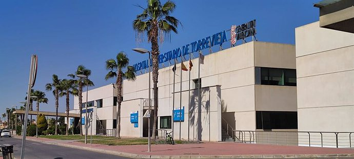
ALICANTE, 22 Mar. (EUROPA PRESS) -
A team of surgeons from the University Hospital of Torrevieja (Alicante) has performed "successfully and in a pioneering manner" a laparoscopic esophageal resection for tumor, with the support of three-dimensional (3D) technology.
The incorporation of 3D technology in a personalized way increases the success rate in complex surgeries. In fact, the surgical team led by Andrés Tomás and integrated, among other members, by Yannko González, has successfully performed this operation on an elderly patient with a tumor in the esophagus, as reported by the Generalitat in a statement.
The patient came to the center with difficulty eating, after several months of evolution and 10 percent weight loss. The personalized 3D anatomical model has made decision-making and performing this surgery easier. Specifically, it has helped the surgical team to locate the tumor "more precisely" and "significantly" improve its oncological resection.
"3D technology has been of great help in countless surgical interventions, although to date, its systematic application in the field of esophageal and gastric surgery is yet to be discovered. We are motivated to become one of the leading hospitals in the application of this technology, in a pathology as exciting and complex as esophageal-gastric cancer," explained Dr. González.
More than 400 scientific publications support the clinical use of 3D modeling technology in personalized cancer surgery planning. These studies highlight the use of individual patient-specific anatomical models to help decrease intraoperative complications in cancer surgery and contribute to increased success in interventions.
Multidisciplinary teams that include engineers, mathematicians, physicists, as well as medical imaging technicians and radiologists work on the development of 3D models. The work process begins when these professionals anonymously receive medical images and radiology reports through an online platform. Using artificial intelligence and advanced computational techniques, they proceed to examine and determine the relevant anatomical structures.
The final 3D model is provided through a specialized platform that facilitates the planning of surgical interventions. In addition, there is the possibility that surgeons request a physical replica of the model, which is made using 3D printing techniques.